
King Scallop Pecten maximus. Painted thorny oyster, Spondylus pictorum. Green snail, Turbo marmoratus. Close up of spire of Strawberry Top Shell, Clanculus pharaonius. Shell pigments responsible for colour have been identified for Clanculus pharaonius (Williams et al. 2016) and Turbo marmoratus (Tixier, 1952) and to class for Spondylus pictorum (Wade et al. 2019) and Pecten maximus (Williams et al. in prep.). Pigments for Clanculus pharaonius and Pecten maximus have also been linked to a molecular pathway (Williams et al. 2017; Williams et al. in prep) images © Natural History Museum.
The evolutionary history of colour and vision has been closely intertwined over the last 500 million years, leading to a dazzling diversity of colours and visual systems in marine invertebrates.
Evolution of colour in the tropical starfish, Linckia laevigata
Heritable colour polymorphisms, where alternate colour morphs occur within a single species and are genetically controlled, provide an unparalleled opportunity to rigorously assess the role colour plays in the evolution of species. However, until now, there was little possibility of investigating colour polymorphisms in marine invertebrates as the pigments responsible for colours in this group are rarely known, and even less is known about their synthesis or inheritance.
The iconic seastar Linckia laevigata is a rare exception to this rule. With its attractive appearance and rainbow of colour morphs, this seastar is a ‘poster child’ for tropical reefs in the Indian and Pacific Oceans. It occurs in various colours, including yellow, orange, grey and purple, but is best known for its royal-blue morph. Exceptionally, the pigments responsible for the iconic blue colouration of the seastar Linckia laevigata have not only been identified as carotenoproteins, but we have obtained full-length sequences of the gene responsible for the protein moiety.
In a study funded by the Natural History Museum and National Geographic, we are determining whether there is a link between variation in this gene and visible colour. Our data will provide the opportunity to further investigate the effect of colour on adaptation, ecology, geographical range and speciation in Linckia and more widely in other species.
Future studies funded by a Catalyst Grant will investigate the role of colour in more detail in Linckia laevigata, with a focus on the blue pigment.
-
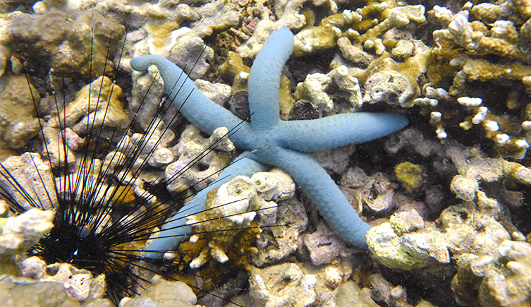
Light blue, Sabah © Nadia Santadomingo
-
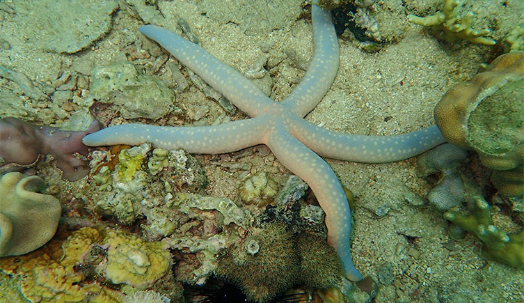
Grey-blue (aboral surface), apricot (oral surface - slightly exposed along edges of arms), Sabah © Nadia Santadomingo
-
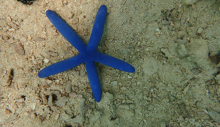
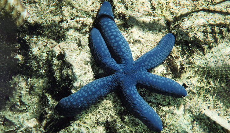
Blue aboral surface, Sabah © Nadia Santadomingo
-
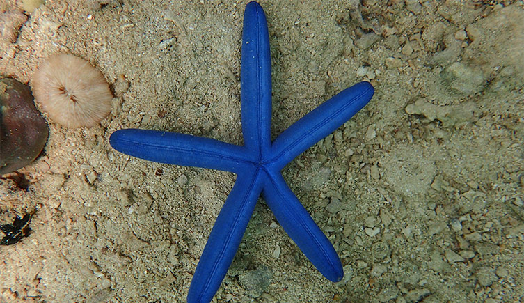
Blue oral surface, Sabah © Nadia Santadomingo
-
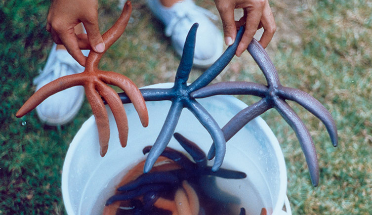
Orange, blue and purple morphs, Thailand © John Benzie
-
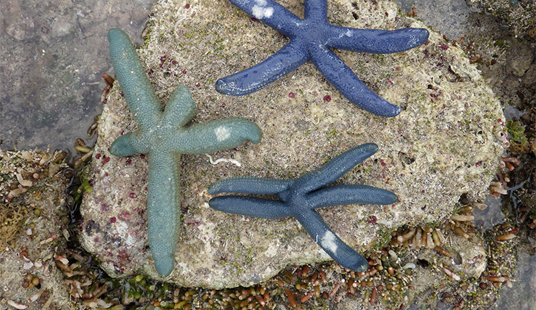
Green-blue and blue aboral surface of three sea stars, Japan © Yasunori Kano
-
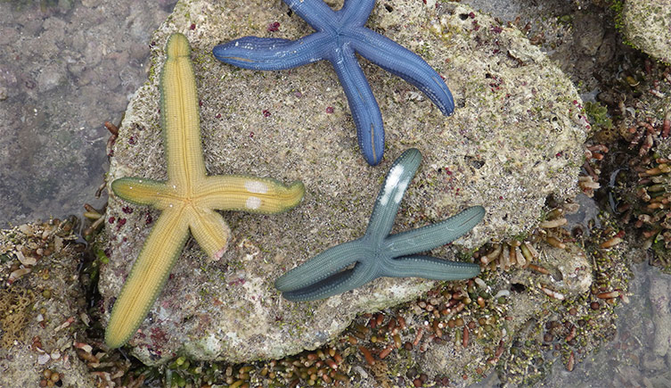
Yellow, blue and green-blue oral surfaces of the same three sea stars, Japan © Yasunori Kano
-
Colour and marine invertebrates
The fabulous and diverse colours and patterns of molluscan shells and brightly coloured echinoderms are widely recognised, however their function for the animal is sometimes less clear and has been the focus of many ecological and evolutionary studies. Despite these studies, almost nothing is known about the evolution of colour in these groups.
My lab is working with international collaborators to identify shell pigments in a range of molluscan species using methods such as high-performance liquid chromatography and resonance Raman spectroscopy.
We are building on that information to identify the molecular pathways responsible for the biosynthesis of pigments in molluscs and echinoderms using transcriptomics and differential gene expression.

Raman studies have shown that partially methylated carotenoids are responsible for these shell colours in the scallop genus Mimachlamys (Wade et al. 2019) images © Natural History Museum
Publications – evolution of colour
Wade, J., Pugh, H., Nightingale, J., Kim, J. S. and S. T. Williams. (2019). Colour in bivalve shells: Using resonance Raman spectroscopy to compare pigments at different phylogenetic levels. Journal of Raman Spectroscopy 50: 1527–1536.
Grant, E. H. & S. T. Williams. (2018). Phylogenetic distribution of shell colour in Bivalvia (Mollusca). Biological Journal of the Linnean Society 125: 377–391. doi.org/10.1093/biolinnean/bly122
Williams, S. T., Lockyer, A. E., Dyal, P., Nakano, N., Churchill, C. K. C. and D. I. Speiser. (2017). Colourful seashells: Identification of haem pathway genes associated with the synthesis of porphyrin shell colour in marine snails. Ecology and Evolution 7: 10379-10397.
Williams, S. T. (2017). Molluscan shell colour. Biological Reviews 92: 1039-1058.
Williams, S. T., Ito, S., Wakamatsu, K., Goral, T., Edwards, N. P., Wogelius, R. A., Henkel, T., de Oliveira, L. F. C., Maia, L. F., Strekopytov, S., Jeffries, T., Speiser, D. I. and J. T. Marsden. (2016). Identification of shell colour pigments in marine snails Clanculus pharaonius and C. margaritarius (Trochoidea; Gastropoda). PLoS One 11(7): e0156664
Williams S.T., J.A.H. Benzie. (1998). Evidence of a biogeographic break between populations of a high dispersal starfish, congruent regions within the Indo-West Pacific defined by colour morphs, mtDNA and allozyme data. Evolution 52: 87-99

Green mussel, Perna viridis ©NHM
Collaborators
National Geographic Funding
Dr Nathan Kenny
Dr Hugh Carter
Dr Chris Laumer
Dr Sumaitt Putchakarn
Mr Surapong Banchongmanee
The Royal Society of New Zealand Catalyst Funding
Dr Nathan Kenny
Dr Yasunori Kano
Dr Karen Cheney
Dr Matthias Fellner
Prof. Miles Lamare
Prof. Maria Byrne
Other Collaborators
Dr Lauren Sumner-Rooney
Dr Jessica Wade
Dr Dan Speiser
Prof. Nick Roberts
Ms Stephanie Heyworth
Lab members
Current Lab Members
Dr Abigail Ingram (Royal Society funded Daphne Jackson Fellow)
Caleb Trimble (PhD Student, University of Otago/NHM)
Karolina Zarzyczny (NERC INSPIRE PhD Student, Southampton University/NHM)
Previous Lab Members
Hugh Carter (NERC London PhD Student, University College London/NHM, 2023)
Alison Irwin (NERC GW4+ PhD Student, Bristol University/NHM, 2022)
Matilde Lanzini (NHM/Imperial College MRes Student, 2021)
Emily Noone (NHM/Imperial College MSc Student, 2019)
Sophie Sykes (NHM/University College London MRes Student, 2019)
Edward Wort (NERC INSPIRE PhD Student, Southampton University/NHM, 2018)
Hazel Pugh (NHM/ Imperial College MRes Student, 2017)
Heather Grant (NHM/ Imperial College MRes Student, 2017)
Dr Nathan Kenny (NHM PDRA, 2016)
Chris Hughes (NHM/IC MSci Student, 2014)
Lisa-Marie Braun (NHM/IC MRes Student, 2012)
Martine Claremont (NHM/Imperial College, MRes Student, 2007; PhD Student, 2011)
Brendan Hever (NHM/IC MSci Student, 2004)
Molluscan vision

Vision in shelled molluscs, like the landsnail on the left, is usually poor. However, our studies are showing that conch shells, like Lentigo lentiginosus, right, image ©David Massemin, have surprisingly good vision for marine snails and we are investigating using CT scanning to look at the eye in more detail.
Nearly all molluscs are thought to be colour blind and most have poor visual acuity, yet they showcase the greatest diversity of eye types in the animal kingdom.
My lab is working to investigate the loss of eyes in dark environments, in particular the deep-sea family Solariellidae (with Dr Lauren Sumner-Rooney).
While it may come as no surprise to find that some solariellids living in the deep sea have lost their eyes, it is surprising that vision has been lost at least seven times in this family of detritovores.
Even more surprisingly, their pigment-cup eyes have degenerated via more than one evolutionary pathway (loss of pigment, degeneration of vitreous body obstruction of aperture), making solariellids an ideal system to look at repeated evolution of vision loss.
My lab has also started looking at the evolution and diversification of eyes in the iconic gastropod superfamily Stromboidea.

We are investigating using CT scanning to look at the eye in more detail (Sumner-Rooney et al. 2019; Irwin et al. in prep) image © Alison Irwin
Publications – molluscan vision
Irwin, A. R., Williams, S. T., Speiser, D. I., & Roberts, N. W. (2022). The marine gastropod Conomurex luhuanus (Strombidae) has high-resolution spatial vision and eyes with complex retinas. Journal of Experimental Biology, 225(16), jeb243927.
Williams, S. T., Noone, E. S., Smith, L. M., & Sumner‐Rooney, L. (2022). Evolutionary loss of shell pigmentation, pattern, and eye structure in deep‐sea snails in the dysphotic zone. Evolution.
Sumner-Rooney, L., Kenny, N. J., Ahmed, F. and S. T. Williams. (2019). The utility of micro-computed tomography for the non-destructive study of eye microstructure in snails. Scientific Reports 9: 15411.
Sumner-Rooney, L. H., Sigwart, J. D., Smith, L. and S. T. Williams. (2016). Repeated eye reduction events reveal multiple pathways to degeneration in a family of marine snails. Evolution 70(10): 2268-2295.

Image © Michael Bok
Watch as Conomurex luhuanus displays changes in behaviour in response to a looming stimulus.
See Irwin et al. (2022) Journal of Experimental Biology, 225(16), jeb243927 for more details.
Read more
-
 Science news
Getting a good look at deep-sea snails
These snails are losing their eyes. CT scanning is uncovering why.
18 December 2019 -
 Oceans
What does it take to study the ocean?
Much of the ocean remains unexplored, but scientists at the Museum are helping unravel the mysteries of the world beneath the waves.
-
 News
NewsWhy sea snails are pretty in pink
Museum-led research uncovers the pigments that give the sea snails Clanculus pharaonius and C. margaritarius their striking pink and yellow-brown shells.
13 July 2016 -


Art and science reflections on colour
Join a group of scientists and artists as they reflect on colour and vision in nature and beyond, how our individual experiences differ, and the impact of technology.


Invertebrate division
Invertebrates are extraordinarily diverse, accounting for 95% of animal species. They can be found almost everywhere, including some of the most extreme environments on Earth.