Create a list of articles to read later. You will be able to access your list from any article in Discover.
You don't have any saved articles.

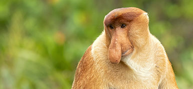
A rare photo of a living solariellid, Ilanga laevissima. The fringe of small tentacles at the front of the animal (around its mouth) move along the ocean floor to pick up sediments and search for food. The long tentacles coming from the head and the sides can detect chemicals in the water and objects by touch. The small, black spot is its eye. © David Herbert
Snails and other molluscs have a huge range of eye shapes and sizes, from tiny pigment cup eyes in limpets, to concave mirror eyes in scallops.
CT scanning is allowing researchers to find out more about this fascinating group of animals.
Animals living in dark environments (such as caves, the deep ocean and underground burrows) can lose their eyes and skin colour. They don't need eyes in the dark, and the challenges associated with living in these environments, for example the increased pressure in the deep sea, mean their energy may be better spent elsewhere.
Vision loss in animals is perhaps most researched in Mexican cavefish and burrowing mammals such as mole rats.
Solariellidae, a group of deep-sea snails classed as gastropods, are also evolving in such a way that over time the eyes of some species no longer function. What makes solariellids unique is that non-functional eyes have evolved multiple times, independently and in multiple ways.
Museum researcher Dr Suzanne Williams is investigating this as part of an ongoing collaboration with Dr Lauren Sumner-Rooney at University of Oxford, and with coauthors Dr Nathan Kenny and Farah Ahmed.
'There are at least three different ways these deep-sea snails are losing their eyes that we know of,' explains Suzanne. 'Their eyes are either shrinking and losing pigmentation, withdrawing into the eye stalks so light can't get in, or experiencing degradation of the vitreous body, which acts as a lens.'
Solariellidae are a family of tiny deep-sea snails ranging from 0.5 to 10 millimetres. They are commonly found between 200 and 1,000 metres on the continental slope, but can be found in water more than 4,000 metres deep. Their size and the difficulty in obtaining them from such locations make them a rare find in most museum collections.

The shell of Elaphriella wareni, one of the three specimens used in the research. In this species, the eyes (not shown here) were covered by the external tissue, sealing the retinal cup. The vitreous body, which may act as a lens in a functional eye, appeared fragmented.
The traditional method of studying these specimens is called histology. This can be time-consuming and destructive as it involves slicing samples into very thin sections and staining them.
But newer technologies are revealing more about these snails than ever before.
CT scanning is a non-invasive and non-destructive technique that uses X-rays to create 3D models of the internal and external features of specimens. This is important for samples that are rare, difficult to collect or part of a collection with limited access due to its historical or taxonomic value.
'If you only have one specimen in the world of something, you're not going to want someone to chop it up,' says Suzanne. 'CT scanning allows us to look at the anatomical features without destroying valuable specimens.'

This sea snail, Bathymophila diadema was collected live from water about 900m deep. The black spot is the eye which has been engulfed by the surrounding tissue. © Lauren Sumner-Rooney
Suzanne used CT scanning to examine the internal anatomy of the snail eyes and found several things that traditional techniques had missed.
The CT scans showed that while the snails were losing their eyes, their energy was being put into other nerves, including mechanoreceptors (responsible for detecting touch) and chemoreceptors (responsible for detecting chemicals). The energy investment could be seen in two long cephalic tentacles on the head and a fringe of small oral tentacles around the mouth.
'We were able to see that there was a lot more energy investment in the nerves going to the tentacles around the head and mouth,' says Suzanne. 'If they can't see, then it's useful that more energy is being put into touch and smell.'
Suzanne also found that the eyes had two nerves coming out of the eye stalk. This contrasted with previous studies that identified only a single nerve in some gastropods, although the two nerves shared a joint origin in the brain.
'The nerves coming from the brain are connected to the eye in two places, not just one,' she explains. 'This is something else we weren't able to see with traditional histology.'

The shell of an Ilanga navakaensis. Found in shallower waters than where other deep-sea snails live, it has open pigment cup eyes located at the top of the eye stalks.
Despite the size of the animals, Suzanne still had plenty to study. The non-destructive approach meant there was no disruption, distortion or damage to the surrounding tissue. This allows Suzanne and future scientists to draw more data from a single specimen as well as a larger area of interest.
The scans provided high-res images, taking only a few hours to complete. This encouraged Suzanne to use CT scanning to take quick measurements of the eyes as well, which proved to be far more accurate than any other way.
'We've been sticking them in the scanner to measure the shape and size of the lenses,' says Suzanne. 'It's precise and saves time.'
The downside to CT scanning is that it cannot identify pigment cells, which are apparent when using traditional histology. Fortunately, the presence of pigment is usually obvious from a quick examination under a dissecting microscope.
Suzanne's research used specimens from the Paris Muséum national d'Histoire naturelle's collection, which had been analysed using traditional histology in 2005-2006. The specimens were Ilanga navakaensis, Elaphriella wareni and Bathymophila diadema.
The chosen specimens were of similar age and had previously been processed in the same way after collection. This is the first time direct comparisons had been made between the two techniques in gastropods.
'CT scanning is one of the most important developments in recent decades when it comes to studying the structure of living things,' says Suzanne. 'The use of CT scanning in the study of eyes stands to gain much from non-destructive tomographic imaging, as resolution, speed and accessibility improve.
Suzanne will continue researching gastropod eyes using CT scanning. She anticipates that as the technique continues to improve and become more economical, it will be used a lot more in future with other studies.
Just how weird can the natural world be?

Discover how comparing dragonfly and human vision can give us a new appreciation for both.

Our oceans are changing fast. Find out how Museum collections are helping scientists to understand the future of marine life.

Meet the creatures with the most crafty, strange and sophisticated eyes in the animal kingdom.

Discover one of the more unusual colourful specimens at the Museum: a colour-changing marvel that transformed with the seasons.