Create a list of articles to read later. You will be able to access your list from any article in Discover.
You don't have any saved articles.

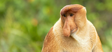
Not much is known about the lives and biology of spectacled porpoises. A specimen that arrived at the Museum in 2017 may provide some answers. © Alona K via Shutterstock
Spectacled porpoises are thought to live in the remote oceanic region surrounding Antarctica.
Little is known about the lives of these animals, so Museum scientists are endeavouring to learn all they can from the dissection of a female that stranded in the Falklands.
This page contains graphic images of dissection that some readers may find upsetting.
In January 2017, a mature female spectacled porpoise (Phocoena dioptrica) was found at San Carlos in the Falkland Islands. This species is so rare that a team from Falklands Conservation promptly froze the animal and contacted the Museum's Principal Curator of Mammals, Richard Sabin.
The specimen was kindly shipped to the UK aboard a British Antarctic Survey research vessel and now offers a valuable opportunity for scientific study as an addition to the Museum's Cetacea research collections. This spectacled porpoise is only the third to be acquired by the Museum, the last having been acquired in 1939.

The spectacled porpoise specimen just hours after being found stranded in the Falkland Islands © Andy Stanworth/Falklands Conservation
Richard put together a team of Museum and external experts to gather as much scientific information about the animal as possible.
Dr Anne-Claire Fabre and Dr Anthony Herrel, experts in anatomy, were invited to lead the initial dissection of the 192-centimetre-long spectacled porpoise. Focusing on an examination of the animal's musculature, their work left the internal organs and skeleton intact to be studied in more detail.
The dissection team worked with care to not damage the skeleton or organs as the latter were removed. These were weighed, photographed and prepared for examination in the Museum's wet lab.
Throughout the dissection process, Museum mammals curators Roberto Portela Miguez and Louise Tomsett also collected samples from the specimen for the Museum's frozen tissue collection.

The spectacled porpoise's organs were removed to be examined for parasites, prey and plastics
With the organs removed, an investigation into their contents began. Scientists were looking for parasites and evidence of the porpoise's diet, as well as possible ingestion of plastics.
Although the specimen initially seemed clear of internal parasites, cetacean parasitologist Dr Natalia Fraija was able to find a number of hair-thin, translucent nematodes (roundworms) in the lungs and liver. Further investigation is needed to determine the species.

A large number of fly eggs were found on the porpoise during a surface examination in 2017. During the dissection, the scientists also searched for parasites inside the body.
On opening the stomach, it became clear that the apparently underweight porpoise hadn't eaten recently prior to death.
Richard, who coordinated the various strands of the investigation, says, 'My suspicion about the nutritional state of the animal was confirmed. When we opened the stomach, it was empty. The only thing we found in the forestomach was the remains of a tiny crab which has been passed to a colleague for identification.'
It's unclear whether the crab was a prey item or accidentally ingested.

Dr Natalia Fraija investigates parasites removed from the spectacled porpoise's organs
Although there were no other visible remains of food items in the stomach or intestines, Richard was able to collect several fluid samples from the forestomach and digestive tract. These will be analysed at the Museum for environmental DNA (eDNA) and will hopefully provide evidence of what this animal may once have eaten.
Alex McGoran is a London NERC DTP PhD student based at Royal Holloway (University of London) and the Museum. Alex primarily studies the presence of plastics in River Thames-dwelling fishes, but has also looked at the stomach contents of seals. Alex was also on hand to help investigate the spectacled porpoise.
'I mostly work on fish, so it's a new experience working on such big animals,' says Alex. 'There were differences in the structure of the digestive tract and stomach between the seal and the porpoise: seals just have one stomach cavity, whereas the porpoise has three before the intestines.'

Nematodes collected from the spectacled porpoise's internal organs
Alex kept the stomach and intestines separate to look at whether more plastics accumulate in one or the other. Multiple stomach cavities mean more opportunities for plastics to get stuck.
'In the porpoise the stomach lining was incredibly folded, which could be really interesting in terms of plastics,' says Alex. 'It could catch all sorts of stuff. So it would be interesting to see if there is more plastic in the stomach than the intestines.'
Finding plastics in the porpoise specimen would provide more evidence on just how pervasive the man-made substance is. But as the animal hadn't eaten recently prior to death, Alex wasn't expecting to find a lot of plastic.

Alex McGoran searches through the intestines for plastics.
'Anything that it does have will be its baseline contamination - just what it gets as it swims. That in itself is quite interesting because if it isn't feeding and is still encountering plastic, then we know that feeding isn't the only pathway for plastic intake.
'We know that plastics can be in the most remote regions. The Antarctic is supposed to be pristine, considering it has the circumpolar currents, but plastic is still being recovered there. So I wouldn't be surprised if we do find plastic, even with how remote the species is.'
Other investigations carried out include Richard and Travis Park, a cetacean evolution researcher, taking a closer look at the head by carrying out a micro-CT scan. This scan can be used to look in more detail at the anatomy of the soft and hard tissues, allowing for comparative studies with other species of porpoises.
Anthony and Anne-Claire are also interested in investigating how the musculature of the upper body connects to the head, to provide insights into locomotion.

Richard Sabin investigates the spectacled porpoise's liver for parasites
At 70 kilogrammes, the spectacled porpoise took three days to fully defrost before dissection could begin.
First to be removed were the skin and blubber layers. This involved precise work to delicately separate the blubber from the muscles beneath.

Dr Anthony Herrel and Dr Anne-Claire Fabre began the dissection by removing the blubber
When the porpoise stranded, staff from Falklands Conservation noted that the animal looked underweight. This was confirmed with the removal of the blubber layer, which was noticeably thin for an animal that is believed to live in the cold waters surrounding Antarctica.
The scientists now know the porpoise's emaciated state was likely from malnourishment. From a first look through the stomach and intestines, Alex didn't find any large pieces of plastic. Large plastics can create a false feeling of fullness, causing animals to stop feeding. Alex's plastics search is now centring on microplastics.

Blubber helps insulate marine mammals in their cool, watery world. This animals's blubber was unusually thin for an species that lives primarily in waters surrounding Antarctica.
Amy Scott-Murray, a 3D imaging specialist from the Museum's Imaging and Analysis Centre, has been recording virtual 3D models of the spectacled porpoise specimen. These models will become a lasting archive of the porpoise's shape, size and appearance.
Amy first carried out a 3D-structured light scan during the specimen's scientific debut in October 2017. Also attending this dissection, Amy scanned the porpoise for a second time after the removal of the skin and blubber.

Amy Scott-Murray began the 3D-structured light scan of the spectacled porpoise once the blubber was removed
This scanner works by projecting a grid pattern onto an object and using cameras to measure how the pattern deforms as it is stretched across the three-dimensional surface. The system then uses a series of dots placed on and around the specimen, which allow the computer to work out where Amy is scanning. These dots are automatically removed from the resulting scan.
This specimen is offering scientists a rare insight into the life and biology of the rarely seen spectacled porpoise. The data and specimens collected - organs, parasites, DNA and skeleton - will be used for research on this species for many years to come.
Richard says, 'We've completed the external examination and the dissection, we've examined all of the internal organs, we found parasites on the outside and inside the animal, we've taken eDNA samples. We've done everything that we set out to do so far and we've had the bonus of also looking for plastics.
'This project is really demonstrating the value of having so much expertise in the Museum, the importance of our international contacts and use of modern analytical technologies.'

Read more about the pioneering work of the Museum's marine scientists.
Just how weird can the natural world be?

The Museum's spectacled porpoise specimen went on a rather unusual trip, all in the name of science.

The spectacled porpoise is one of the least understood cetaceans of all, so there is a lot to learn from the Museum's new specimen.

Little is known about the lives of spectacled porpoises. A rare find is allowing Museum experts to find out more.